I. Esophageal Cancer
Types
Adenocarcinoma (lower third)
Squamous cell carcinoma (upper two-thirds)
Risk Factors
Adenocarcinoma: GERD → Barrett’s esophagus (intestinal metaplasia → dysplasia → adenocarcinoma)
Squamous: Smoking, alcohol, achalasia, caustic ingestion
Paraneoplastic Syndrome
Hypercalcemia (PTHrP secretion by squamous cell)
Diagnosis
Initial: Barium swallow (optional if dysphagia + alarm signs absent)
Best: EGD with biopsy
Staging: Endoscopic ultrasound (EUS), CT chest/abdomen
Treatment
Resectable: Esophagectomy
Locally advanced: Neoadjuvant chemoradiation → surgery
Metastatic: Palliative chemo + esophageal stent for obstruction
II. Gastric Cancer
Types
Adenocarcinoma
Intestinal type (better prognosis, older patients)
Diffuse type (signet ring cells, younger patients)
Risk Factors
H. pylori
Smoking
Nitrosamines
Chronic atrophic gastritis, pernicious anemia
Blood type A
EBV infection (associated with diffuse type)
Paraneoplastic Associations
Acanthosis nigricans
Leser-Trélat sign (sudden seborrheic keratosis eruption)
Diagnosis
EGD with multiple biopsies (>6)
Staging with EUS, CT, diagnostic laparoscopy (for peritoneal metastases)
Treatment
Surgery (subtotal or total gastrectomy)
Neoadjuvant chemotherapy if T2 or higher stage
HER2-positive tumors: Trastuzumab + chemotherapy
III. Small Bowel Cancers
Types
Adenocarcinoma (especially duodenum)
Carcinoid tumor (ileum > appendix > rectum)
Lymphoma (especially ileal)
GIST
Risk Factors
Crohn’s disease → adenocarcinoma
Celiac disease → enteropathy-associated T-cell lymphoma
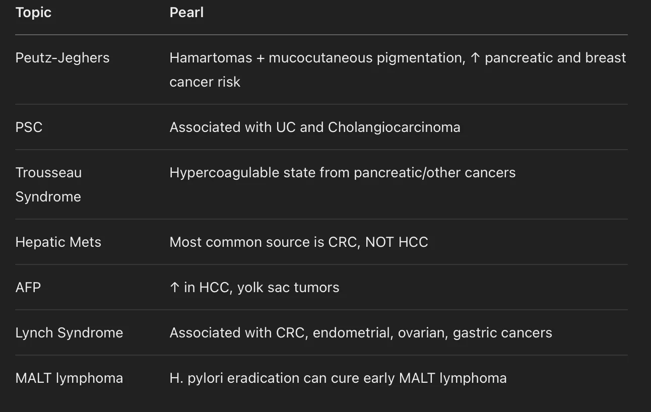
Peutz-Jeghers syndrome
Diagnosis
Capsule endoscopy
Double-balloon enteroscopy
Biopsy for confirmation
Treatment
Adenocarcinoma: Surgical resection
Carcinoid: Surgical resection if >2 cm or at base of appendix
GIST: Resection + Imatinib for metastatic disease (c-KIT mutation)
IV. Colorectal Cancer
Types
Adenocarcinoma (arises from adenomatous polyps)
Genetic Syndromes
FAP (APC mutation, 100% CRC risk)
Lynch syndrome (MLH1, MSH2 mutations)
Associated cancers: Endometrial, Ovarian, Gastric
MUTYH-associated polyposis
Juvenile polyposis
Peutz-Jeghers (hamartomatous polyps)
Screening Guidelines
Average risk: Start colonoscopy at age 45, every 10 years
1st-degree relative <60y or 2 relatives any age: Start at 40 or 10 years before earliest case, every 5 years
Lynch: Start at 20-25 years, every 1-2 years
Clinical Presentation
Right-sided: Iron deficiency anemia, fatigue
Left-sided: Obstruction, "apple core" lesion
Paraneoplastic
Streptococcus bovis (gallolyticus) bacteremia → screen for CRC
Diagnosis
Colonoscopy + biopsy
CEA (monitoring)
Treatment
Surgery
Chemotherapy for Stage III (node-positive) and high-risk Stage II
Rectal cancer: Neoadjuvant chemoradiation preferred
V. Anal Cancer
Type
Squamous cell carcinoma
Risk Factors
HPV 16, 18
Immunosuppression (HIV)
Smoking
Diagnosis
Biopsy
Anal Pap smear in HIV-positive patients
Treatment
Nigro protocol: Chemoradiation (5-FU + Mitomycin C + radiation)
Surgery reserved for recurrence
VI. Hepatocellular Carcinoma (HCC)
Risk Factors
Hepatitis B (especially perinatally acquired)
Hepatitis C
Alcohol cirrhosis
Hemochromatosis, alpha-1 antitrypsin deficiency, NASH
Aflatoxin (Aspergillus)
Paraneoplastic
Hypoglycemia (tumor consuming glucose)
Erythrocytosis (EPO production)
Screening
US + AFP every 6 months in cirrhotics and HBV patients
Diagnosis
Imaging (triphasic CT or MRI): Arterial enhancement + venous washout
No biopsy needed if classic imaging!
Treatment
Resect small tumors with preserved liver function
Liver transplant (Milan criteria)
TACE, RFA for inoperable tumors
Sorafenib for advanced/metastatic disease
VII. Cholangiocarcinoma
Risk Factors
Primary sclerosing cholangitis (PSC)
Liver flukes (Clonorchis sinensis, Opisthorchis viverrini)
Biliary tract stones (choledocholithiasis)
Diagnosis
MRCP preferred
ERCP + brushings/biopsy
Treatment
Surgery (if resectable)
Palliative chemotherapy if unresectable
VIII. Gallbladder Cancer
Risk Factors
Gallstones
Porcelain gallbladder
Chronic Salmonella (typhi) infection
Diagnosis
Ultrasound initially
CT for staging
Treatment
Cholecystectomy if early
Poor prognosis once metastatic
IX. Pancreatic Adenocarcinoma
Risk Factors
Smoking (strongest)
Chronic pancreatitis
Diabetes mellitus (new-onset after age 50 = red flag)
Obesity
Family history
Genetic Syndromes
Peutz-Jeghers syndrome
Lynch syndrome
BRCA1/2
MEN1
Clinical Presentation
Painless jaundice (head tumors)
Courvoisier’s sign (non-tender enlarged gallbladder)
Migratory thrombophlebitis (Trousseau sign)
Depression
Weight loss, anorexia
Diagnosis
First test: CT scan with pancreas protocol
Best confirmatory: Endoscopic ultrasound (EUS) + biopsy
CA 19-9 elevated (used for prognosis, not diagnosis)
Treatment
Whipple procedure (pancreaticoduodenectomy) if localized to head without metastasis
Chemotherapy (FOLFIRINOX, Gemcitabine)
Palliative biliary stenting for obstruction
⚡ Step 2 CK Special High-Yield Reminders
🛎 Quick Shelf Tips:
Progressive dysphagia → Think esophageal cancer (start with solids).
Painless jaundice → Think pancreatic head cancer or cholangiocarcinoma.
Right-sided CRC → Iron deficiency anemia in an older adult.
Left-sided CRC → "Apple core" lesion on imaging.
H. pylori → MALT → Adenocarcinoma sequence if untreated.
AFP elevated in HCC (not diagnostic alone).
Always biopsy to confirm cancer (unless classic imaging like HCC multiphasic CT/MRI).
✉️ Stay Ahead in Your Medical Journey!
Want more high-yield study guides, exam-crushing strategies, and exclusive updates for the NBME Shelf and Step 2 CK?
👉 Join our free email list and get:
Early access to new mind maps, flowcharts, and study tools
Weekly high-yield tips tailored for med students
Special bonus: a downloadable Medicine Shelf Rapid Recall Guide when you sign up!
🔵 Join Now — It’s Free!
Newsletter Block This newsletter signup form needs a storage option. Edit the block and enter a storage location via the Storage tab.